


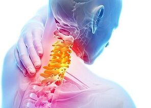
Osteochondrosis of the cervical spine (ICD code – 10 – M42) ranks second in frequency of cases, after injury to the intervertebral discs in the lumbar region.High daily load or, conversely, low physical activity cause negative changes in vertebral structures.
What signs are characteristic of cervical osteochondrosis?How dangerous is compression and destruction of intervertebral discs?What are the symptoms and treatment, prevention methods, consequences of an untreated disease?
Causes
Deformation of the vertebral structures of the cervical spine is a consequence of the action of negative factors and pathological processes:
- poor posture;
- disturbances in metabolic processes due to poor nutrition;
- spinal injury;
- wear of vertebral discs during the natural aging of structures in old age;
- genetic predisposition to orthopedic diseases;
- physical inactivity, prolonged computer use, causing tension in the neck muscles;
- development of an infectious-inflammatory process due to hypothermia, exposure to drafts;
- defects in the development of vertebral structures in the cervical region.
Factors that provoke damage to the vertebrae, cartilaginous tissue and discs:
- obesity;
- very intense training;
- the head is thrown back or lowered to the chest for a long time during work;
- flat feet;
- incorrect body position when working while sitting;
- frequent microtraumas of the cervical spine due to very intense training.
First signs and symptoms
At the initial stage of the disease, the patient rarely associates frequent headaches, discomfort in the shoulders, numbness in the neck during prolonged stay in a stationary position with damage to the vertebral structures.Most of the time, there is no effective treatment and the pathology progresses.

In the second to fourth stages of osteochondrosis, negative manifestations in the cervical region are most pronounced.In advanced cases, negative signs spread to other parts of the body: hearing aid, eyes, upper limbs.
Doctors identify several types of characteristic symptoms:
- root.Pain syndrome in the parietal, occipital region, neck, discomfort in the back muscles, shoulder girdle.An important point is the loss of shoulder muscle reflexes.Radicular syndrome is most often observed in the third or fourth stage of the pathology, significant impairment of the condition of the vertebrae and discs and a noticeable decrease in the height of the bone structure;
- muscle tonic.Painful sensations appear in the chest and heart region.Acute symptoms usually occur in the inner arm muscles;
- neurovascular.When the capillaries and vertebral artery are compressed, the patient complains of headaches and dizziness.
Development stages:
- first.Under the influence of negative factors, small cracks appear in the intervertebral discs, the integrity of the nucleus pulposus is disturbed, fluid gradually leaks, a decrease in elasticity is observed and the disc shrinks.A slight cracking sound is heard in the vertebrae when moving the head;pain and numbness in the cervical region are disturbing when the body is in one position;
- second.The pathology progresses, a violation of the integrity, ruptures and a decrease in the density and thickness of the fibrous ring appear.The result of negative processes: squeezing the core into the resulting cracks and breaks.The patient's condition worsens and performance decreases.The strength of the headaches increases, stiffness appears in the cervical spine, the face, neck and arms become numb;
- third.Bulging of the disc, active compression of the structure, loss of elasticity lead to protrusion and hernia of the intervertebral disc.Neurological disorders appear, a rupture of the fibrous ring occurs, the clinical picture is clearly expressed: reflex syndrome and radicular syndrome are worrying.Many patients complain of weakness in the hands, fingers, decreased hearing and vision acuity;
- room.In advanced cases, the discs become flattened, images show the growth of osteophytes and compensatory phenomena develop to maintain the spine in its usual position.The pain syndrome weakens somewhat, but the stiffness of the cervical spine remains.
Diagnosis

The appearance of pain in the cervical region, frequent dizziness, decreased mobility and sensitivity of the fingers, the development of other signs of deforming spondylosis are a reason to consult a vertebrologist, neurologist or orthopedic traumatologist.The doctor finds out the clinical picture, lifestyle, diet, history of spinal injuries, chronic diseases.It is important to understand the causes of osteochondrosis and identify the factors that cause degenerative changes in the discs between the vertebrae.
Next, the specialist prescribes an examination:
- tomography – computed tomography and magnetic resonance imaging;
- Dopplerography of blood vessels is carried out in advanced stages, the presence of pronounced neurological disorders, frequent fainting, numbness in the shoulder area, hands and fingers;
- X-ray of the cervical spine (in the initial stages of pathology, the method is not very informative).
Effective treatments
In the early stages of the development of deforming spondylosis in the cervical region, special gymnastics and external agents with analgesic and anti-inflammatory effects are sufficient.The patient must monitor his posture.You should not stay in the same position for a long time, warm up every hour and do neck exercises daily.
As the pathology progresses, local remedies and gymnastics are not enough to eliminate the complex of negative symptoms.The degenerative-dystrophic nature of the changes affects the mobility of the cervical region, the pain syndrome intensifies, protrusions and herniated discs appear, doctors identify all types of radicular symptoms and reflexes of deforming spondylosis.
Important!Only an integrated approach to therapy combined with cessation of smoking, alcohol and dietary changes can partially or completely correct the situation.It is important to eat well, include foods rich in calcium, phosphorus, fresh vegetables, cheese and drink plenty of fluids.In the absence of timely and competent therapy, there is a risk of the patient's disability.
Medicines
The doctor selects medications to eliminate negative manifestations of various types:

- fight against pain syndrome.Names with analgesic and anti-inflammatory effects are effective.The best option is NSAIDs in the form of tablets, injections, gels and ointments.Additionally, you can take painkillers prescribed by your doctor;
- elimination of swelling.Fluid retention in tissues is eliminated by diuretics, dehydrating compounds and hormonal medications.Take medications in this group strictly as prescribed by your doctor: powerful diuretics can cause serious heart and blood pressure problems;
- chondroprotectors to restore cartilage quality.When the elastic tissue is damaged, friction between adjacent vertebrae increases, the density of the layer decreases and bulging of the disc and rupture of the fibrous ring are possible due to compression of the vertebrae.Doctors prescribe medications with a combined effect.The formulations contain not only glucosamine and chondroitin, but also anti-inflammatory ingredients.Various multicomponent products provide a good effect;
- increased blood circulation in problematic vertebral structures.The doctor selects medications that thin the blood and compounds that dilate blood vessels;
- fight against muscle spasm.Against the background of weakened bone structures and dilapidated cartilaginous tissue, muscles try to maintain the correct position of the spine;Excessive overexertion causes pronounced pain.To relax tissues, muscle relaxants are prescribed;
- activation of metabolism, increased local immunity.The patient takes vitamin and mineral complexes, dietary supplements with calcium, biostimulants and antioxidants.
Physiotherapeutic procedures
Increasing the impact on problem areas, activating blood circulation, delivering drug components to the affected area, nutrition, reducing inflammation - this is not a complete list of the beneficial properties of physiotherapy sessions.The doctor prescribes a set of procedures in addition to taking medications and therapeutic exercises.
Effective types of influence:
- laser therapy;
- darsonval;
- drug electrophoresis;
- diadynamic currents;
- infrared radiation;
- electrical stimulation of the affected muscles.
Therapeutic gymnastics
With osteochondrosis, it is important to maintain the mobility of the cervical vertebrae and strengthen the muscles and ligaments.After examining and clarifying the condition of the affected structures, the doctor prescribes special exercises for the cervical spine.Simple actions maintain the elasticity and flexibility of structures.

Therapeutic exercises for neck muscles:
- turn your head to the left and then to the right;
- head tilt with gradual increase in amplitude, forward and backward;
- circular movements.At first, the amplitude is minimal, gradually increasing the “circle”, but without the appearance of pronounced crushing of the cervical vertebrae.
Perform all movements smoothly, shaking is prohibited.During exercise, you should not allow your vertebrae to grind too much or cause sharp pain.The amplitude must be increased gradually, otherwise injuries to the cervical spine are possible.Simple but effective exercises should be performed not only during therapy, but also to prevent osteochondrosis.The frequency of classes is two to three times a day, the duration of gymnastics is no more than five to seven minutes.Doctors recommend mandatory and daily implementation of the complex for people who have a “sedentary” job.
Manual therapy
Doctors are cautious about realigning the cervical vertebrae.Most experts do not recommend manual therapy for osteochondrosis of this area.The slightest deviations during the procedure, more sudden movements, incorrect application of the technique - and the session can end in paralysis or fracture of the cervical vertebrae with serious consequences, including death.
Osteochondrosis becomes “younger” every year.To avoid damage to bone and elastic structures, you need to pay daily attention to an important part of the spine, perform simple exercises, change body position more often during the work process, not smoke and eat well.If characteristic signs of spondylosis deformans appear, you should contact an orthopedic traumatologist or vertebrologist.